

Vascular Tunic Of The Eye
Beefcake and function of the heart
Author: Maria Yiallouros, Erstellt am 2016/eleven/21, Editor: Maria Yiallouros, English language Translation: PD Dr. med. Gesche Tallen, Final modification: 2016/xi/21 https://kinderkrebsinfo.de/doi/e182894
Table of contents
- Layers of the center
- The inner office of the eyeball
- How the eye works
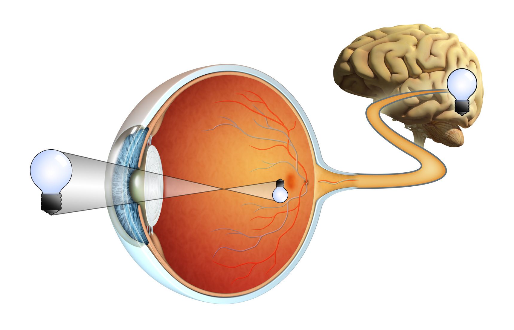
The eye is a sensory organ. It collects light from the visible world effectually us and converts information technology into nerve impulses. The optic nerve transmits these signals to the encephalon, which forms an epitome then thereby providing sight.
Man eyes primarily consist of two world-shaped structures, the eyeballs, which are surrounded past the the bony sockets of the skull, the orbits. The orbits are covered with fatty and fibrous tissue to protect the eye. Additional structures protecting the eye include the eyelids, the outer coating layer of the centre (fibrous tunic), the conjunctiva, and the lacrimal glands. Six special muscles that insert at dissimilar sites outside the eyeball work together to command centre movement.
Each eyeball houses the following parts of the middle:
- the three coating layers: the outer, middle and inner coat
- the inner function of the eyeball: it contains the lens and the vitreous body and is divided into the anterior and the posterior sleeping accommodation.
The following chapters volition explicate beefcake and function of the three coats equally well as of the inner part of the eyeball.
Layers of the middle
The eyeball is surrounded by a three-layered wall, the three coats of the eye. They consist of different tissue and serve unlike functions.
Outer glaze (fibrous tunic)
The center's outer layer is made of dense connective tissue, which protects the eyeball and maintains its shape. It is as well known as the fibrous tunic.
The gristly tunic is composed of the sclera and the cornea. The sclera covers nearly the entire surface of the eyeball. With its external surface being white-coloured, information technology is commonly known as the "white of the center". The sclera provides attachments for the muscles that control the eye'south movement (meet to a higher place).
The transparent cornea occupies the front middle function of the external tunic. Information technology serves as the middle's "window", which lets the lite in and bends its rays, thereby providing most of the eye'south focusing power.
The anterior, visible part of the sclera too as the inner surface of the eyelids are covered by the conjunctiva, a mucous membrane that helps lubricating the centre together with the tears made by the lacrimal glands, thus protecting the eye from drying out.
Heart coat (vascular tunic)
The eye layer of tissue surrounding the eye, likewise known as the vascular tunic or „uvea", is formed – from backside forward – by the choroid, the ciliary body, and the iris.
The choroid takes up the posterior v-sixths of the bulb and is mainly comprised of blood vessels. Its major functions are oxygen supply and nutrition for the center. A nighttime pigment, melanin, occurs throughout the choroid in club to help limiting uncontrolled reflection within the eye, which would potentially result in the perception of confusing images.
The inductive role of the choroid passes into the ciliary body, 1 part of which is anchoring the lens in place. The ciliary body contains a muscle (ciliary muscle), which tin change the shape of the lens for adjustment to far or near sight, respectively, thereby decision-making the so-called refractive power of the lens (accomodation). Additional functions of the ciliary body are the production, secretion, and outflow of aquaeous sense of humor (the latter via the so-called „Schlemm'southward canal"), a watery fluid that fills both the anterior and the posterior chambers of the middle (see beneath).
The iris, which is connected to the anterior office of the ciliary body, covers the meridian of the lens. Like to the aperture of a camera, information technology controls how much calorie-free is let into the middle. The iris forms a round, thin structure within the eyeball that regulates the size and the diameter of the student. Information technology too contains pigments, the amount of which determines a person's eye colour. For example, in children with blue eyes, the iris contains less pigment than in brownish-eyed kids.
Inner coat
The third and inner coat of the middle is the retina, which is responsible for the perception of images – vision.
The retina is a light-sensitive layer of nervous tissue composed of multiple sensory cells, then-chosen light- or photoreceptor cells, equally well as associated nerve cells and other types of cells, all working together to brand a person meet.
For vision, there are two types of photoreceptor cells: rods and cones. Rods provide the perception of black-and-white vision, mostly in dim light, whereas cones help to see colors in daylight.
The light and colour impulses received by these photoreceptors are transmitted to the associated nerve cells of the retina, which, on their part, send these signals – via the optical nerve – to the visual middle (visual cortex) of the encephalon.
The point where the optic nervus fibers depart from the eyeball (optical disc) does non contain any photosensitive cells; it is, thus, insensitive to low-cal and termed the "blind spot".
Directly opposite the lens, the retina contains a small yellowish area, the "macula lutea". Its central part (fovea centralis) is densely packed with cone cells for color perception. At this indicate, the sense of vision is the virtually authentic and detailed.
The inner part of the eyeball
The inner part of the eyeball consists of the lens, the vitreous body and the two eye chambers.
The lens
The lens is a transparent olive-shaped structure in the heart that has no claret vessels. Lens and cornea (come across above) piece of work together to focus the light rays passing through the eyeball to the back of the centre, that is, to the retina, by bending or refracting them, thereby creating clear images of the surround perceived from different distances.
By adjusting its shape and size, the lens tin can alter the focus. This procedure is called accomodation. Accomodation is possible thank you to the lens' elastic capsule as well as to the lens fibers, which connect with the ciliary muscle (see heart layer of the center).
The vitreous body (vitreous humor, vitreous)
The vitreous is a clear gelatinous mass held by collagen fibers. It is situated betwixt lens and retina and comprises near 2 thirds of the unabridged eyeball. By pushing the retina towards the choroid, the vitreous promotes keeping the retina in place.
Anterior and posterior centre chamber
The anterior bedchamber of the eye is located between the iris and the cornea (see in a higher place). The posterior chamber is the infinite between parts of the iris and the lens. Both chambers are filled with aquaeous fluid to attend cornea and lens.
How the eye works
The human eye is a circuitous optical organisation that basically works like a photographic camera: the iris serves every bit the aperture that controls the amount of light rays reaching cornea and lens (photographic objective), and the retina works as the film.
(© Andrea Danti - Fotolia.com)
Bending of lite rays by cornea and lens serves to create abrupt images on the retina. These images ultimately trigger nervus impulses, which are transmitted to the encephalon where the images are perceived and interpreted.

Vascular Tunic Of The Eye,
Source: https://www.gpoh.de/kinderkrebsinfo/content/diseases/solid_tumours/pohretino_patinfo120120611/the_eye/index_eng.html
Posted by: mcmullenalliat.blogspot.com
0 Response to "Vascular Tunic Of The Eye"
Post a Comment